


Next: Deconvolution
Up: Basics in Imaging, Deconvolution
Previous: Weighting
The Miriad task invert
has been configured for the reduction
of SMA data. The maximum number of spectral windows has been extended
to a limit of 48. Miriadtask invert
forms images from visibilities. Both continuum
images or spectral line cubes can be formed using invert. It
can generate images or image cubes for several polarizations, as well
as handling multi-frequency synthesis and mosaicing observations. Miriadtask
invert
can also form complex-valued images from non-Hermitian
data (e.g. holography data). Appropriate point-spread functions
(dirty beams) can also be generated.
invert% inp
  Task:   invert
  vis      = sgra-star.cal  % uvdata contain 24 chunk-
                                 averaged spectral points.
  map      = sgra-star.map     % name of dirt images
  beam     = sgra-star.beam    % name of dirt beams
  imsize   = 512,512           % size of the image
  cell     = .15               % cell size
  sup      = 0                 % weighting function; 0
                                 for natural weighting
  stokes   = xx
  options  = mfs,sdb,systemp   % using mfs, and produces
                                 spectral beams and normal
                                 beams; weighting by the
                                 data variance.
Fig. 4.1 shows the dirty image of this data. Because of the discrete
sampling function, the image is contaminated by the side lobes of the
point spread function (Fig. 4.2). Apparently, a point source is
dominant in the image (Fig. 4.1).
Figure:
Fourier transform of the weighted visibility data or a dirty map.
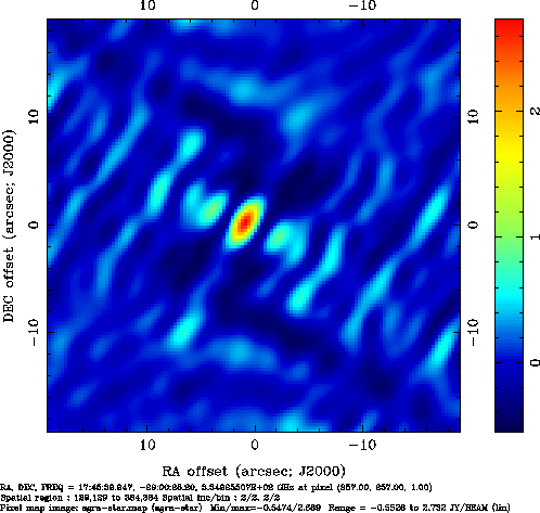 |
The side lobes must be minimized. There are a number of algorithms
that can deconvolve the dirty beam from the dirty map.
Figure:
The point spread function or dirty beam of the visibility data.
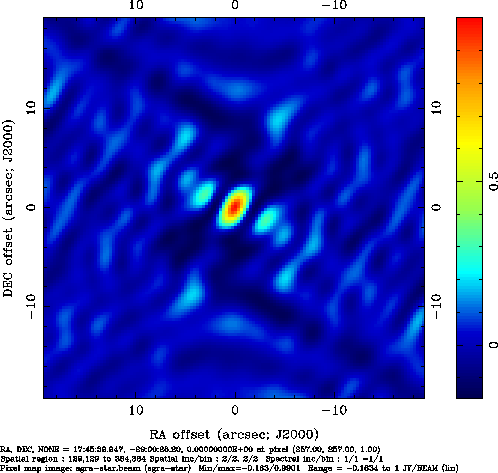 |
Next: Deconvolution
Up: Basics in Imaging, Deconvolution
Previous: Weighting
Jun-Hui Zhao (miriad for SMA)
2012-07-09